Author(s):
Edelhoff, D., Prandtner, O., Saeidi Pour, R., Liebermann, A. , Stimmelmayr, M., Güth. J.-F.
Publication Date:
2018-02
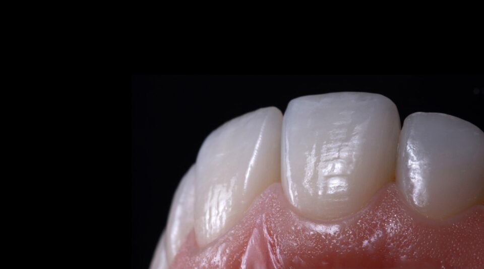
DownloadAnterior tooth restorations: The performance of ceramic veneers.
The establishment of the adhesive techniques in combination with tooth-coloured restorative materials is among the greatest achievements in restorative dentistry.
Adhesively luted veneers based on various silicate ceramic materials have made a major contribution to this. Ceramic veneers, which for a long time had the nimbus of purely aesthetic restorations, are still being used for a growing number of indications. They have developed into a serious treatment alternative to classical, far more invasive forms of restoration. Today, they are used in addition to numerous other applications to reconstruct the biomechanics of teeth. They can also be used to set an appropriate function and even mask heavily discoloured endodontically treated teeth.
The article describes the principles of modern veneering technology using clinical examples, with special emphasis on the cooperation with the dental technician in the analysis, definition of the treatment objective, colour determination, decision in favour of a suitable ceramic, preparation design and determination of the luting material for adhesive placement. In addition, the long-term behaviour of ceramic veneers is evaluated on the basis of the existing scientific literature.
Analysis and determination of the restorative objective
The analysis of function, aesthetics and biomechanics as well as the definition of the restoration goal requires very close communication with the dental technician. The following documents should be available for the laboratory analysis for fabricating the diagnostic wax-up:
- Portrait photographs frontally and laterally with and without facebow
- Recordings with slightly opened mouth and relaxed upper lip as well as video recordings (smartphone is sufficient)
- Recordings in which the patient speaks freely and counts once, for example, from “50” to “55
- intraoral photos of both jaws (photo status)
These images support the determination of the length and inclination of the maxillary middle incisors. This is the central starting point of any anterior tooth- or more complex rehabilitation. Furthermore, impressions of both jaws showing the vestibulum including the jugae alveolaria and a jaw relation determination are required.
Make sure to also check out our relating treatment concepts and video-tutorials!